New stem cell mechanism in your gut EurekAlert!
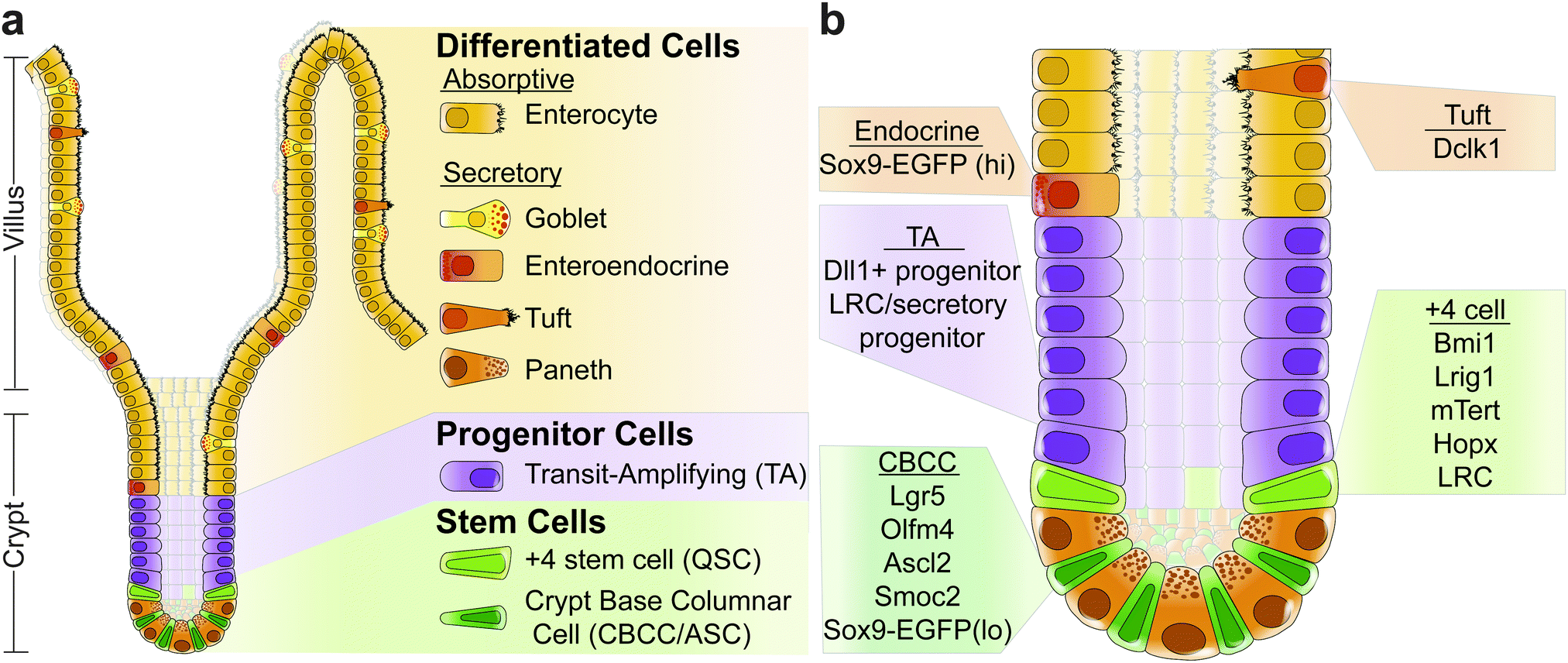
Crypt-villus structure and continuous proliferation enable the intestine to act as an absorptive organ and a protective barrier. Tissue replenishment is fuelled by adult stem cells that divide.

Does the crypts of lieberkuhn also consists of microvilli which is present in the villi as the
The surface of the small intestine is lined by a monolayer of tightly packed, polarized epithelial cells organized into invaginations called crypts, and finger-like protrusions called villi 1,.

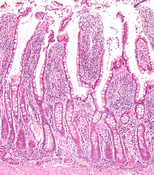
Histological aspects of the duodenal villi and crypts for the control... Download Scientific
The initial crypt formation process occurs by a symmetry-breaking event; the crypt-villus separation process is also recapitulated in a 3D organoid culture system. When a crypt-like protrusion is generated from a round-shaped organoid, specialized wedge-shaped cells referred to as hinge cells appear at the crypt/villus boundaries ( Sumigray et.
Artwork Showing Structure Of Small Intestine Villi Photograph by John Bavosi
In the intestinal epithelium, proliferated epithelial cells ascend the crypts and villi and shed at the villus tips into the gut lumen. In this study, we theoretically investigate the roles of the villi on cell turnover. We present a stochastic model that focuses on the duration over which cells mig.

Intestinal structure (A) diagram of small intestine showing crypts,... Download Scientific
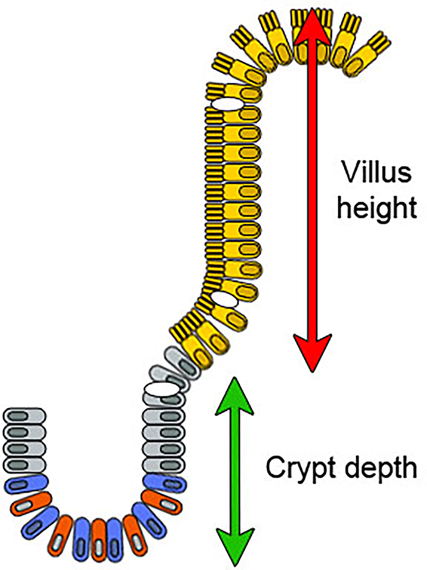
Intestinal crypts and villi develop in human fetuses from 8-24 weeks gestation with an increase in crypt depth and villus height with advancing gestational age 1.
Cells and Their Association With Crohn's Disease Owlcation
Intestinal crypts, which are composed of stem cells (SCs) and Paneth cells, are an essential unit for epithelial homeostasis 1. In both mice and humans, crypts form after the emergence of.

Determination of the villi/crypt ratio. The villi/crypt ratio (V/C... Download Scientific Diagram
Several in vitro models have been developed to study the intestinal epithelium. (a) Organoids are 3D organotypic cultures obtained from dissociated intestinal crypts, in which cells self-organize, with an enrichment of stem cells in crypt-like domains and of differentiated cells in villi-like regions along the central lumen. (b) 2D self.
5 Schematic of duodenal villi and crypts. Download Scientific Diagram
The adult mammalian intestine is composed of two connected structures, the absorptive villi and the crypts, which house progenitor cells. Mouse crypts develop postnatally and are the architectural unit of the stem cell niche, yet the pathways that drive their formation are not known.
Frontiers Apolipoprotein A4 Defines the VillusCrypt Border in Duodenal Specimens for Celiac
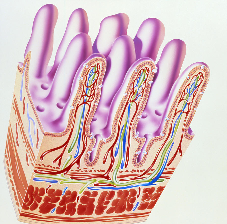
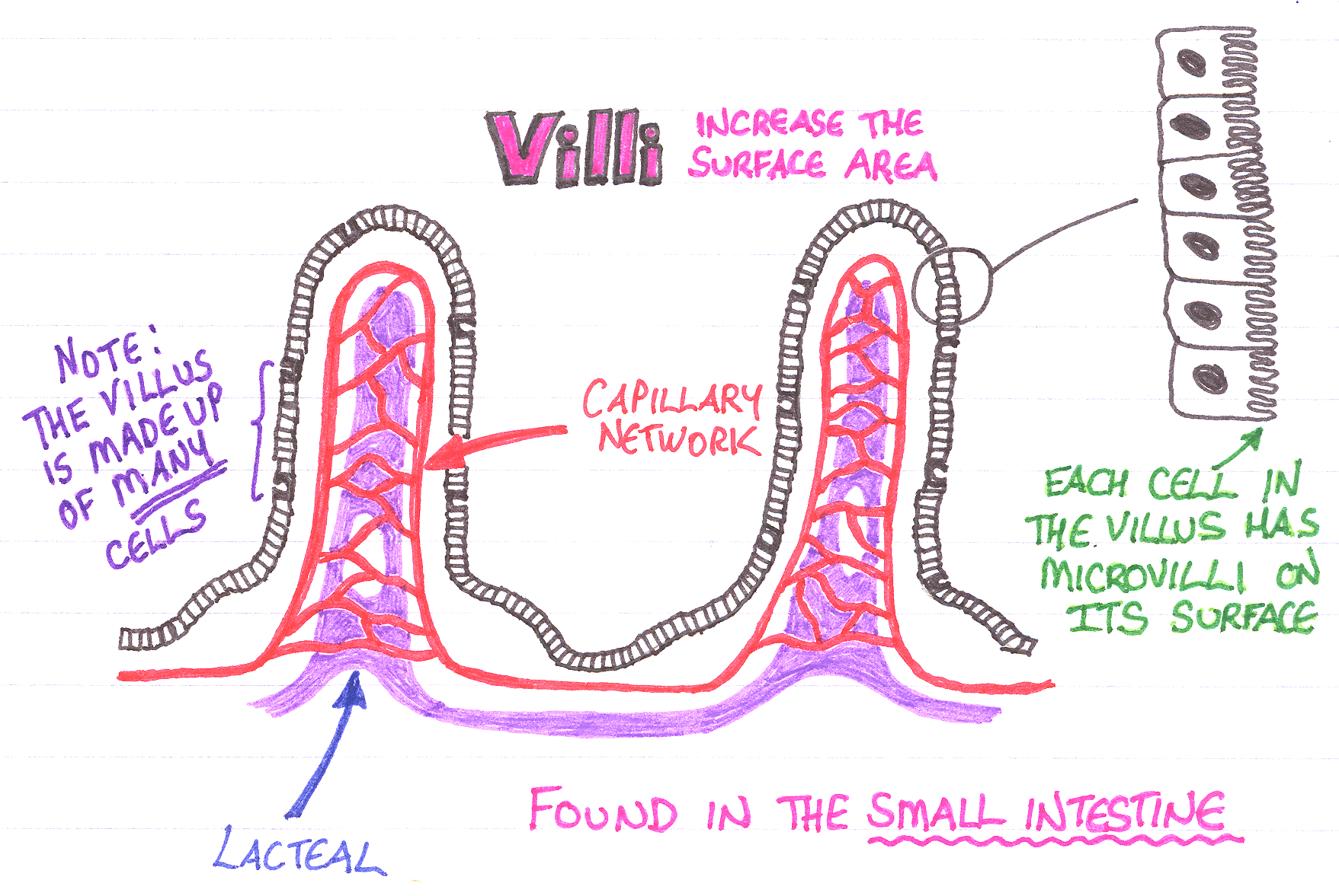
Between the villi there are crypts, called crypts of Lieberkuhn, which extend down to the muscularis mucosae. These crypts are short glands. The lamina propria which underlies the epithelium has a rich vascular and lymphatic network, which absorbs the digestive products, and there is a muscularis mucosae layer immediately at the base of the crypts.
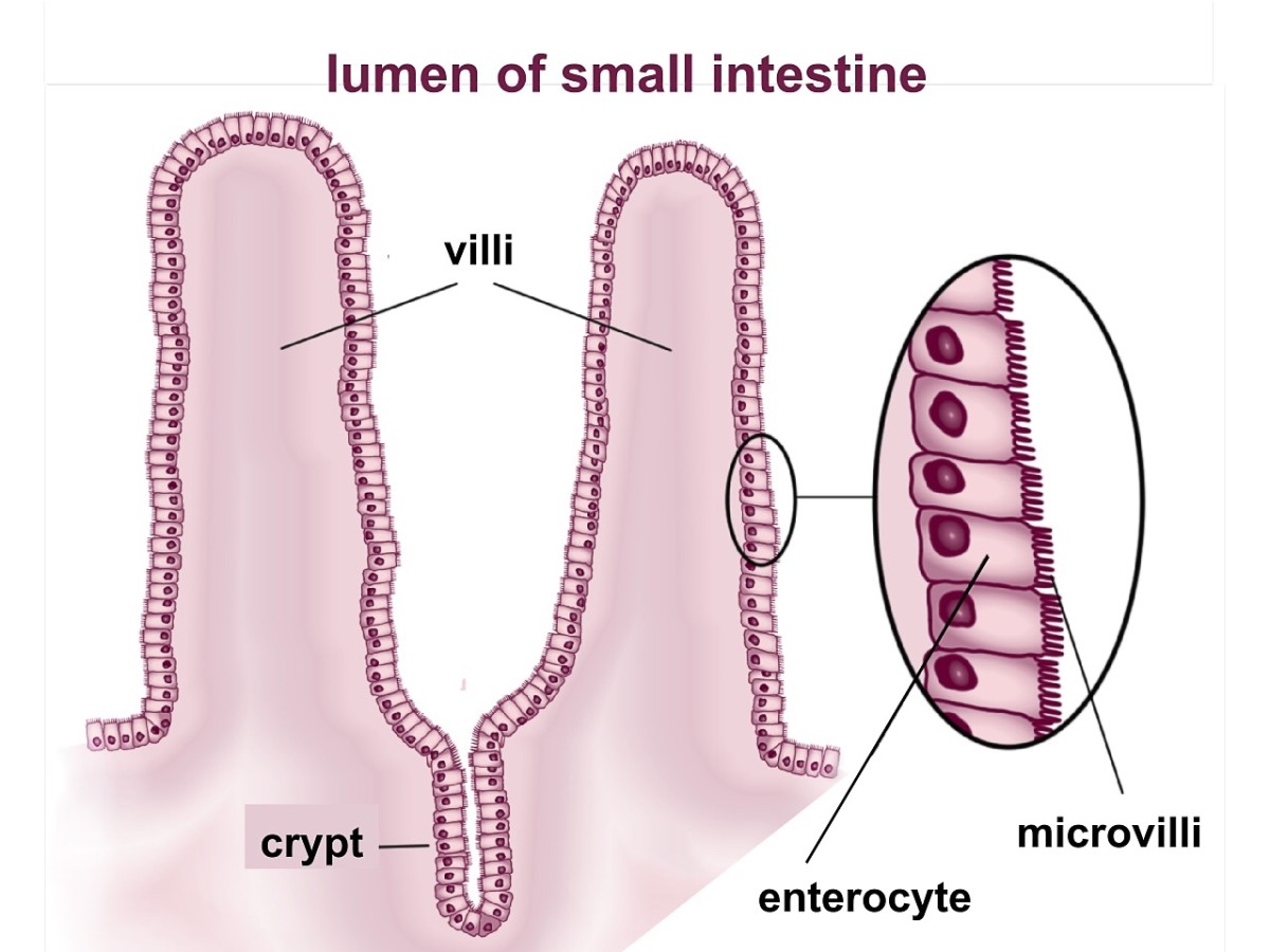
What is the Difference Between Villi and Microvilli
This layer is organised into finger-like protrusions (villi), which serve as the site of nutrient absorption and pockets (crypts) containing mostly proliferating cells. This outer layer of.
Unraveling intestinal stem cell behavior with models of crypt dynamics Integrative Biology
Crypts only begin to emerge in the small intestine after villus morphogenesis is complete. Epithelial cells located in the inter-villus regions undergo myosin-II-driven apical constriction, which.

Cellular architecture of small intestinal and colonic epithelial... Download Scientific Diagram
In histology, an intestinal gland (also crypt of Lieberkühn and intestinal crypt) is a gland found in between villi in the intestinal epithelium lining of the small intestine and large intestine (or colon).
:max_bytes(150000):strip_icc()/GettyImages-103018448-01b8ff85b5a243588652ac05c6bff972.jpg)
How the Intestinal Villi Help With Digestion
The inner lining of the gut consists of a single cell layer of intestinal epithelium that forms millions of crypts and villi. Stem cells (shown in green) reside at the bottom of the crypts and replicate daily, generating new cells to maintain the tissue. Image courtesy of Unmesh Jadhav/HMS, DFCI
Why do we have villi in the small intestine? Socratic
The mesenchymally-derived basement membrane dynamically controls morphogenesis, cell differentiation and polarity, while also providing the structural basis for villi, crypts and the microvasculature of the lamina propria so that tissue morphology, crucially, is preserved in the absence of epithelium.

Diagram Of Villi In Small Intestine diagramwirings
The epithelium of the small intestine is organized into large numbers of self-renewing crypt-villus units. Villi are finger-like protrusions of the gut wall that project into the gut lumen to maximize available absorptive surface area.

Pictures A&B Gastrointestinal villus showing the crypt, enterocyte, and microvilli Wenger Feeds
The villi limit epithelial cells from shedding early or remaining in the epithelium for long periods by separating the villus tips, where cells shed, from the crypts, where cells are produced. Here, we propose even shedding of epithelial cells as another role of the villi and underline their physiological and pathophysiological importance.